2 Simple Tests for Assessing Ophthalmic Health
Pamela K. Kirby, RVT, Purdue University College of Veterinary Medicine
Schirmer tear testing and fluorescein staining of the cornea provide important information about a patient’s ophthalmic health. (See Schirmer Tear Test & Fluorescein Stain.) The Schirmer tear test (STT) measures basal tear production and reflex tear response and is often used to diagnose keratoconjunctivitis sicca (KCS). cl,ss="intro-deck">1sup> Basal tears are normal tears produced daily to lubricate and nourish the ocular surfaces. cl,ss="intro-deck">2sup>
The STT is performed by inserting an absorbent paper test strip between the lower eyelid and cornea and measuring the length of strip that absorbs moisture in 1 minute. The result is then compared with established normal values.
Fluorescein is a water-soluble stain applied to the cornea to detect corneal ulcers. The stain appears orange in high concentration, such as on the commercial strip, but green when diluted with saline. The stain does not penetrate the intact corneal epithelium but adheres to the hydrophilic stroma when a break in the epithelium occurs.
Indications for Testing
Ophthalmology practices may routinely include Schirmer tear testing in all new patient examinations, but in general practice, testing typically is performed only in patients with signs of ophthalmic disease, including mucoid or crusty discharge, dull corneal surfaces, and prolapsed third eyelid glands.3
Schirmer Tear Test & Fluorescein Stain
Schirmer tear test
The measures basal tear production and reflex tear response and is often used to diagnose keratoconjunctivitis sicca.
Any patient that presents with an ocular problem
Most importantly, patients with
Blepharospasm
Diabetes mellitus
Facial nerve paralysis
Hyperadrenocorticism
Hyperemia
Hypothyroidism
Mucoid discharge
Prolapsed gland of the third eyelid
is a water-soluble stain applied to the cornea to detect corneal ulcers.
Red and/or painful eyes, including those with suspected keratoconjunctivitis sicca
Patients being treated with topical steroids
Canine breeds predisposed to KCS (eg, bulldogs, Lhasa apsos, West Highland white terriers, cocker spaniels) should undergo Schirmer tear testing at every wellness visit.3,4 KCS occurs less commonly in cats,5 so feline patients are not tested routinely. Diagnosing KCS in cats can be difficult because they have sympathetic control over their lacrimal glands and an accurate STT measurement cannot be obtained.6 Patients receiving treatment with sulfonamides should also undergo Schirmer tear testing before and during treatment because sulfonamides have been shown to be cytotoxic to lacrimal gland tissue.4,7 Twenty-five percent of dogs treated with sulfonamides develop KCS.8 Anticholinergic drugs used systemically and topically are also known to transiently decrease tear production.9
In addition to identifying abnormal lacrimation, Schirmer tear testing helps diagnose other ophthalmic conditions. Epiphora, an increase in tear production, is a sign of ocular discomfort. If the STT reading is excessive (ie, >30 mm wetting/minute), causes of ocular discomfort (eg, entropion, ectopic cilia, distichiasis, corneal ulcers) should be explored. Epiphora can also result from a conformational abnormality such as the absence or abnormally small opening of nasolacrimal puncta or medial entropion occluding the nasolacrimal puncta.<sup10 sup>
Proper Patient Restraint
Proper patient restraint helps ensure successful Schirmer tear testing and fluorescein staining. Most patients respond best to minimal restraint. When restraining a dog, place one hand behind the head and one hand under the chin. (See Figure 1.) With cats, place 2 thumbs behind the ears and the index fingers under the jaw. (See Figure 2.)
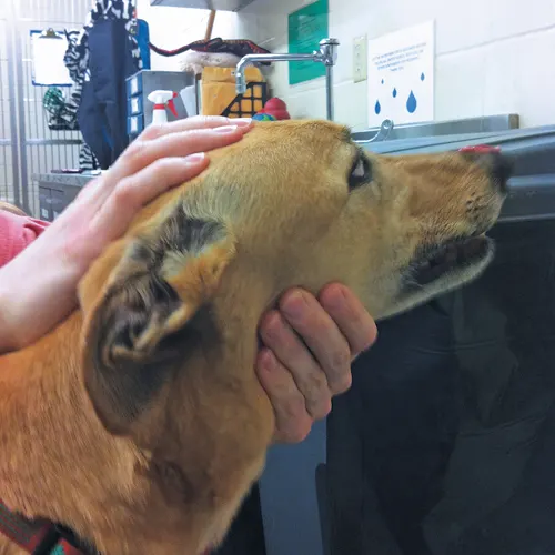
Figure 1 Proper restraint of a dog for Schirmer tear testing and fluorescein staining
Photos courtesy of Pamela K. Kirby, RVT
The Schirmer Tear Test
Perform the STT before instilling any medication in the eye. Clients should be advised to stop giving their pet any ophthalmic medications at least 2 hours before tear measurement.11 Any thick mucus present on the cornea or in the conjunctival sac should be gently removed with a cotton ball or gauze square to prevent the mucus from interfering with the test.
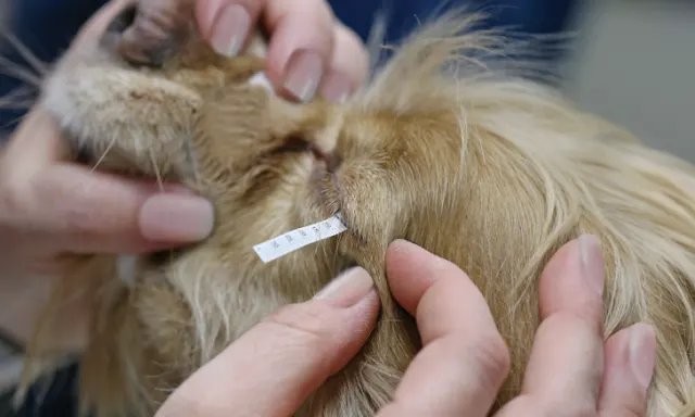
The STT package contains 2 sterile strips. (See Figure 3.) Before opening the package, bend the strip approximately 90° at the notch near the rounded end. Open the package at the opposite end to avoid touching the rounded tips and contaminating the strips with oil from the skin.2 Test each eye separately. Insert the bent, rounded tip of the strip between the lower eyelid and cornea approximately one-half to two-thirds of the eyelid length away from the medial canthus. (See Figure 4.) The strip should not sit between the third eyelid and lower lid. If the patient will not remain still, gently hold the eyelids closed. The strip should be left in place for 1 minute, removed, and read immediately using the scale on the strip. The line at which the moisture stops wicking is the result—moisture may or may not correspond to the blue dye on the strip.
A normal value for dogs is ≥15 mm wetting/minute.10 A 10 to 15 mm wetting/minute result in dogs is considered borderline and treatment should be instituted if the patient is showing clinical signs of KCS. If the patient is not showing any clinical signs, the tear values should be rechecked at the next visit. Results <10 mm wetting/minute in dogs should be considered indicative of KCS and treatment instituted.10
Fluorescein Staining
Always use sterile fluorescein strips (see Figure 5) or single-use sterile vials of fluorescein dye to stain the cornea because multi-use vials are easily contaminated. Moisten a sterile strip with sterile ophthalmic solution (see Figure 6) and touch the strip to the conjunctiva (see Figure 7), or allow the dye to drip from the strip onto the corneal surface. If using single-use vials, allow 1 or 2 drops of the solution to flow over the cornea and into the lower conjunctival fornix.12 To prevent the strip from creating microabrasions on the corneal surface, do not allow the strip to directly touch the cornea. Allow the patient to blink, or gently close and open the eyelids to ensure the cornea is completely covered with the stain and then gently irrigate the eye with sterile ophthalmic solution.
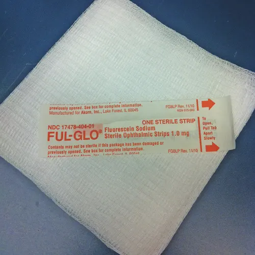
Figure 5 Sterile fluorescein test strips
A break in the epithelial surface appears bright green. (See Figure 8.) Using an ultraviolet or cobalt blue light enhances stain-uptake visualization. Roughened areas of the cornea may appear as a light green haze, indicating corneal scarring from previous injuries, including abrasions caused by STT strips. Light green hazing can also indicate stain is beginning to leak under the loose edges of the epithelium of an indolent ulcer. If a green stain is donut- or halo- shaped with no stain uptake in the center of the circle, a descemetocele should be suspected. Fluorescein stain does not adhere to the lipid-rich Descemet’s membrane.13,14
Conclusion
Schirmer tear testing and fluorescein staining are effective tools for diagnosing ophthalmic disorders in veterinary patients. Patients with a predisposition for KCS should be monitored annually using the STT. Both tests require minimal patient restraint. Prepackaged test strips make performing the tests convenient in specialty and general practices.
When used appropriately, Schirmer tear testing and fluorescein staining are simple, effective tools for diagnosing ophthalmic disorders.
1Take care not to handle the end of the Schirmer tear test strip because oils from the skin can alter the results.
2Touch only the conjunctiva when applying fluorescein stain to the eye to avoid damaging the cornea.
3Ophthalmology practices include testing in all new patient examinations, but general practices should test patients based on history and clinical signs.